Specialists in Companion Animal Neurology (SCAN)
Magnetic Resonance Imaging (MRI)
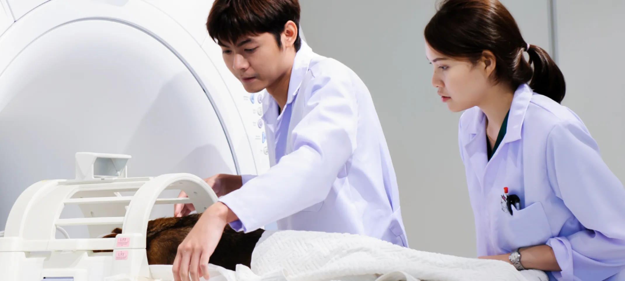
Magnetic resonance imaging, more commonly known as an MRI, is a highly useful tool used in veterinary medicine when more traditional diagnostic measures cannot provide an accurate diagnosis for diseases in the brain or the spinal cord.
Why do we need an MRI?
X-rays are very helpful for evaluating the integrity of the bones of the central nervous system (skull and vertebrae), but unfortunately, the areas where most of the problems occur (brain, spinal cord, intervertebral discs) are not able to be seen with X-rays. MRI is the gold standard imaging technique for the central nervous system in both human and veterinary medicine, allowing us to see the contents inside the bones. Additionally, we can see the anatomy of interest in all 3 image planes, yielding the necessary information to accurately diagnose your pet’s problem.
Is performing MRI harmful to my pet?
No. MRI uses hydrogen atoms present within the body to create an image. There is no ionizing radiation (as there is with X-rays and CT). Bottom line: there are no expected adverse effects to having an MRI performed. The biggest concern associated with MRI is that it needs to be performed under general anesthesia; it is important to know that your pet’s safety is our primary concern. We utilize state-of-the-art monitoring equipment on our patients and one of our highly trained veterinary nurses will be closely monitoring your pet inside the MRI suite at all times.
Why is anesthesia necessary?
Although MRI is incredibly useful for imaging the nervous system, it is also very susceptible to artifacts caused by the movement of the patient. In veterinary patients, the only way to ensure that they lay completely still for minutes at a time is to place them under general anesthesia. The patients are maintained under a light plane of anesthesia (i.e. not a surgical plane) just so they remain still. In this context, a light plane of general anesthesia is safer than heavy sedation as it allows us to ensure appropriate breathing during the procedure.
How does this process work?
After our board-certified neurologist (Dr. Carnes or Dr. Cook) examines your pet, they will discuss where in the central nervous system he/she needs to be imaged in order to evaluate for a “lesion” or problem causing neurologic dysfunction. This will be the primary site that needs to be visualized using MRI.
High-field versus low-field MRI: Does it make a difference?
Yes. Not all MRI machines are created equal. A “low-field” MRI unit has a field strength of less than 1 tesla (T). Any MRI unit with a field strength of 1T or more is considered a “high-field” MRI. The drawbacks of a low-field MRI include 2-3 times longer scan time (meaning more time under anesthesia for your pet), as well as poorer image quality yielding more non-diagnostic scans.
Where is the MRI done?
Specialists in Companion Animal Neurology are the only veterinary practices on the Gulf Coast of Florida that have a high-field MRI located on-site. Other veterinary specialty practices may offer the ability to perform a high-field MRI scan, however, this is performed off-site at a human medical facility. Having an MRI performed off-site may pose several issues including:
Your pet must be transported back and forth from the hospital which can sometimes mean more stress.
It may require additional anesthetic episodes for your pet, as well as a delay in diagnosis and treatment.
The anesthetic and monitoring equipment is often minimal as the facility is designed for human use.
The MRI is not available 24/7 which means when your pet has an emergency an MRI may not be an option.
Having an MRI performed off-site may pose several issues including:
Your pet must be transported back and forth from the hospital which can sometimes mean more stress;
It may require additional anesthetic episodes for your pet, as well as a delay in diagnosis and treatment;
The anesthetic and monitoring equipment is often minimal as the facility is designed for human use;
The MRI is not available 24/7 which means when your pet has an emergency an MRI may not be an option.
What is the cost of an MRI?
The cost varies depending on what area of the body needs to be scanned and how long the scan is expected to take. A complete, written estimate will be presented to you by your technician following examination by the neurologist.
How long will it take and when will we get the results?
In most circumstances, this can be performed on the same day as your pet’s appointment. Most MRI scans for veterinary patients take approximately 30-75 minutes, depending on the complexity and extent of the problem. You will be informed of the results of your pet’s MRI the same day it is performed via an interpretation by one of our Board-Certified Neurologists.

